Friday, June 6, 2014

Vinca Rosea Information-Uses and Benefits

Cape Periwinkle is an evergreen shrub grows throughout varying land of India. Its domestic name is sadabahar or Rose periwinkle. Its botanical name is Vinca Rosea and it belongs from Apocynaceae family. Vinca Rosea is from Plantae kingdom and division is Magnoliophyta which means it is a flowering plant. It is dicotyledons and gentianales in order. The whole plant has medicinal purpose specially the root, seed and the leaf.

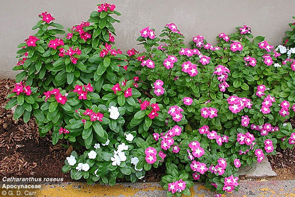
Vinca Rosea is a charming, evergreen shrub, bearing pink or white slaverform flower. It is a native plant of southeastern and eastern Madagascar. After the invasion of European colonist the plant achieved world recognition because of its ornamental property. It can also grow in the tropical and subtropical regions. Vinca Rosea has waxed, glossy covering leaves, which is oval or oblong in shape and oppositely arranged in pairs, with a pale midrib and a short petiole. The flowers usually have five petals like loves.
Vinca Rosea grows very well in infertile and well drained soil but has the chances of dying if the soil is too fertile or healthy. Occasional pinching of the bud is necessary for full growth and branching. It doesn’t need dead heading as the flower drops off after it completes blooming. It is very helpful for the treatment of Leukemia occurring in children. It is prone to the infection of Phytoplasmas which reduces the growth of the leaf.
It is rich with 70 different types of alkaloid specially indole type. Alkaloids like ajamalicine, serpentine and reserpine vinblastine is present in it which has medicinal importance.

  
Cape Periwinkle has n number of uses. For example Chinese used it for herbal medicine, which is used in the treatment of diabetes, Hodgkin’s disease and malaria. The plant is rich in chemical substances like vinblastine and vincristine, which is helpful in the treatment of Leukemia in children and lymphoma. The group of alkaloid which is present in it can be virtually used in the treatment of cancer. The alkaloids may have certain side effects on the body. Traditionally the root bark of priwinklen is used for treatment. It has calming effect and it can also reduce blood pressure and it also contains the alkalois Alstonine.
Some precautionary measure should be taken before its use. Periwinkle is poisonous in nature and restricted for external use like injection or smoking. It has been poisonous for animals grazed on it. It has unwanted side effects after used for the treatment of cancer by doctors. If consumed orally it has hallucinating effect. It is harmfull for kidney and may cause nerve problem. Very low dosage of the plant only under the supervision of doctor is strictly recommended.

Tuesday, June 3, 2014

Valeriana Wallichii Information-Uses and Benefits

Tagara is an amazing sleep-inducing Ayurvedic herb. It is an effective and a powerful natural herb to cure insomnia caused by any reason. Right from the ancient period, Tagara has been known as a superb herb that has a number of beneficial medicinal properties.
The root of the plant is used for preparing different medicines. This is an effective medicine for curing anxiety and nervousness, stress and boosting sleep and making you feel relaxed and fresh. Tagara is very effective in curing insomnia disease in which a person suffers from sleeplessness. This herb relaxes the mind and body and releases all the stress from it. This way it helps in getting a sound sleep and making you feel fresh and relaxed. Tagara is also known as Valerian Wallichi.

There may be several reasons for insomnia. Valerian wallichi is an herb which cures insomnia regardless of the actual reason behind it. It soothes the body and relaxes the mind. This herb is one of the most efficient ways to treat high blood pressure and calming doen the mind. This works as wonder and helps in getting a good sleep. You may take tagara to relax your mind and body and to give instant energy to your body.
Tagara is also very effective in treating skin problems. The paste of tagara leaves may be applied on skin to cure skin problems like- itching, redness, eczema, boils, psoriasis and ulcers. This herb gives a cooling effect which helps in curing these skin problems in an effective way. This helps in making your skin look healthy and clear.

Tagara is also used for reducing pain. This herb is also useful in reducing blood pressure. Tagara can also be used for digestive problems like diarrhea, digestive ulcers and constipation. It helps in cleaning up the digestive system and removes all types of wastes and toxins from your body. It also rejuvenates the body cells which make you look smart and young.
This natural medicine is also useful in curing respiratory infections like asthma, cold, cough and other similar problems. It also cures fever if it is due to any infection. This herb is also good for eyes. Since this herb has cooling effects and calming properties, it not only relaxes mind and body but also act as tonic for the nervous system and eyes. This way, this herb is throughout an effective medicine for many problems.

Monday, June 2, 2014

Tribulus Terrestris Information-Uses and Benefits

Tribulus Terrestris is a herb. It is used as traditional medicine in China and India since time immemorial. It is also called as Puncture vine and only in the mid twentieth century it became known in North America. Eastern European Olympic Athletes had a major role in popularizing it in the West. It improved their performance after using the medicine containing Tribulus.

Steroidal saponins, is the active compound present in Tribulus. Further it has two types, Furostanol glucosides and Spirostanol glucosides , which effects along with tribulus. Saponins are found originally in the leaf.
Tribulus has many uses. People use it for sexual disorders like infertility, erectile problem, low libido and others issues. The last decade is a witness of its popularity in the sport performance. Research performed in Bulgaria and Russia says that Tribulus increases the level of hormones like testosterone, DHEA and estrogen. A more recent study says that one month of Tribulus supplement, 10 to 20 milligrams of per kg body weight, had nil effect in the improvement of male sex hormones, testosterone, androstenedione or leutinizing hormone in comparison to non users of Tribulus.
Early animal studies said that tribulus heightened sexual behavior and intracavernous pressure. This early findings only turned out to be notions for the lack of well designed human studies.
Tribulus is very popular as a sports performance aid but a well designed study proved it wrong. Fifteen subjects were randomly introduced to the dosage of tribulus in 3.21mg per kg body weight regularly. After two months of resistance training there were no changes in body weight, percentage fat, dietary intake, or mood in either group. Most surprising was the improvement in muscle endurance in the placebo group. The placebo group experience improvement in muscle endurance for the bench and leg presses. The tribulus group only experienced an improvement in leg press strength.
Tribulus Terrestris is prescribed to take at a dose between 85 to 250 mg three times daily, with meals. Pregnant or nursing women are prohibited to take tribulus. Gynaecomastia or increase in breast size in a male weight lifter was reported after he went under the dosage of herbal tablets containing tribulus. Breast or Prostrate cancer patient should not take tribulus or those who have hormone-dependent condition should avoid taking it. It has a fatal effect in sheep. It is found to cause a chronic, progressive irreversible disorder in the dopamine circuits of the nigrostriatal complex in the brain. It results into impaired muscle function and weakness in the hindquarters, consequentially leading to death.

Monday, May 19, 2014

Preparing for a Corneal Translant

If you are advised to undergo a corneal transplant, your ophthalmologist will tell you what is required. The transplant will be scheduled according to the condition of your eye and the availability of a donor cornea. Occasionally, a shortage of donated corneas may delay surgery.

If both your eyes need new corneas, the second transplant will not be performed until the first eye has stabilized, which may take up to a year.After surgery, you may be permitted to return home the same day, or you may be requored to stay in the hospital for a day or more.
Some Facts you may like to know:
  • It is not necessary to find a cornea with a matching tissue or blood type.
  • The race, gender, and eye color of the donor are not important.
  • A corneal transplant won’t change your natural eye color.
  • The Cornea heals slowly and improvement in vision may take a year or more.
  • It is difficult to shape the new cornea perfectly. So, astigmatism (a condition where the cornea has an irregular shape, making images seem blurred or distorted) is common after a corneal transplant. However, this can be corrected.

Corneal Transplant

The Cornea is the front, outermost layer of the eye. Just as a window lets light into a room, the cornea lets light into the eye. It also focuses the light passing through it to make images.
Corneal problems can happen to anyone at any age. Sometimes due to disease, injury or infection the cornea becomes cloudy or warped.A damaged cornea, like a frosted or misshapen windowpane, distorts light as it enters the eye. This not only causes distortion in vision, it may also cause pain.
When there is on other remedy, doctors advise a corneal transplant. In this procedure an ophthalmologist surgically replaces the diseased cornea with a healthy one to restore clear vision.

What is Corneal transplant?
A transplant is the replacement of damaged or diseased tissues or organs with healthytissues or organs. In a corneal transplant, the cloudy or warped cornea is replaced with a healthy cornea. If the new cornea heals without problems, there may be tremendous improvement in vision.
The healthy corneal tissue used for transplantation is supplied by an Eye Bank. Eye banks workround the clock to collect, evaluate, and store donated corneas. The corneas are collected from human donors within hours of death. Stringent tests are done to ensure the safety of the person receiving the cornea. The Eye Bank verifies the donor’s medical history and cause of death, and performs blood tests to ensure that the deceased person did not have any contagious disease, such as AIDSor hepatitis.Since the cornea was one of the first parts of the body to be transplanted, corneal transplants remain one of the most common, and most successful, of all transplants.
How does the Eye Work?
Anything you see is an image that enters your eye in the form of light. The different parts of your eye collect this light and send a message to your brain, enabling you to see. For perfect vision all the parts of your eye need to work properly.
  • The cornea is the clear, outer layer of the eye.
  • The pupil is an opening that lets light enter the eye.
  • The iris, the colored paart of the eye, makes the pupil larger or smaller.
  • The lens bends to focus light onto the retina.
  • The retina receives light that has been focused by the cornea and lens.
  • A clear (vitreous) gel fills the inside of the eye, giving it shape.

Wednesday, May 7, 2014

Preparing The Corneal Transplant Surgery

The Corneal Transplant Surgery:
If you are taking any other medication, ask your ophthalmologist whether you should continue it. You will probably be asked not to eat or drink anything for several hours before the surgery.
Usually a local anesthesia is used for surgery, so you will be awake but feel no pain. Intravenous medications will help you relax. The nerves in your eye will be completely numbed so you will not be able to see or move your eye. Sometimes the doctor may use general anesthesia.

The Trnsplant Procedure :
For the transplant, the doctors use an operating microscope and very delicate instruments. Once the old cornea is removed, the new cornea is stitched into place. The sutures or stitches are barely visible and are not painful, althoughyou might feel some irritation or a scratching sensation for a few days.
If necessary, other procedures may be performed at the same time as your transplant. For example, a cataract may be removed and replaced with an intraocular lens(IOL). An IOL may be replaced or removed. The vitreous gel may be removed from the eye and replaced with fluid. A damaged iris may be repaired. Your ophthalmologist will advise you about them.
Some Potential Risks :
As with other surgical procedures, a corneal transplant involves some risks- most of them can be treated.
  • eye infections
  • failure of the donor cornea to function normally
  • rejection of the donor cornea by your body
  • cataract (clouding of the eye’s lens)
  • glaucoma(build-up of fluid, leading to increased pressurein the eye)
  • bleeding from the iris
  • swelling or detachment of the retina
Rejection of a Transplant – the dander signals!
Rejection of a transplanted cornea can occur any time, but is more likely to happen in the first year after surgery. Unfortunately, rejection reduces the chance of success of any repeat corneal transplantation. However, this can be prevented by timely diagnosis and appropriate manaement.
Watch out for these dangersignals:
  • Redness
  • Sensitivity to light
  • Vision loss
  • Pain
The acronym ‘RSVP’ can help you remember these symptoms. If you notice anyof these symptoms in your operated eye, however minor they may seem and regardless of the time of day, contact us immediately. If this is not possible, visit the nearest ophthalmologist, preferble a cornea specialist.

Friday, May 2, 2014

Diabetes and The Eye

Diabetes mellitus or diabetes is characterized by increased levels of blood sugar, due to impaired production or reduced effectiveness of insulin.Patients with diabetes stand the risk of developing diabetic retinopathy due to changes in the blood vessels of the retina caused by poor glucose control. Blindness due to diabetic retinopathy is one of the leading causes of preventable blindness.


The retina is a layer at the back of the eye that senses light and sends images to the brain. In diabetic retinopathy the blood vessels in the retina become fragile and get blocked, leading to water collection (edema), lipid deposition, retina hemorrhages, and the formation of new vessels on the retina. Vision loss can occur due to the lipid and water deposition in the center of the retina (diabetic maculopathy), or due to bleeding inside the eye from new blood vessels(vitreous hemorrhage), or due to membrane formation over the retina which “pulls” on the retina (traction retina detachment).
Symptoms:
In the early stages there are no warning signs; the treatment works best in this stage if detected by a routine retina examination. In relatively advanced stages, the vision worsens gradually or suddenly, making reading or driving difficult. bleeding in the eye an lead to black spots or floaters or total blockage of vision. Persons with advanced diabetic retinopathy may find it difficult to:
  • recognize faces from a distance or read bus numbers,
  • read fine newsprint, bills, or low contrast text,
  • write in a straight line,
  • tolerate bright light or see in dim light,
  • move about independently outdoors after dusk, and
  • tell the time a wristwatch or read the print on an insulin syringe.
Need for early detection
The life expectancy of diabetic patients has increased with the availability of better medicines. However this means an increase in the incidence of diabetic retinopathy and its blinding complications. People with diabetic retinopathy are 25 times more likely to experience permanent vision loss than those with other sight-threatening ailments. Early detection and appropriate treatment can help prevent this. The only method of early detection is a regular and dilated retinal examination.
The recommended check-up schedule for a patient of diabetes with no diabetic retinopathy or its milder forms is:
  • No or minimal retinopathy detected – once a year
  • Mild to moderate non – proliferative diabetic retinopathy – between 6 to 12 months
  • Moderate to severe non – proliferative diabetic retinopathy – between 3 to 6 months
  • Very severe non – proliferative diabetic retinopathy- 2-3 months
  • After photocoagulation – 1 to 6 months or as advised by the treating ophthalmologist.
Diabetic Retinopathy and Pregnancy
Pregnancy can increase the porogression of diabetic retinopathy. Hence pregnant woman with diabetes should have an eye examination every three months. Controlling blood sugar after the progression of diabetic retinopathy has less effect than controlling it in the initial years of the disease.